Open Access
Fig. 6
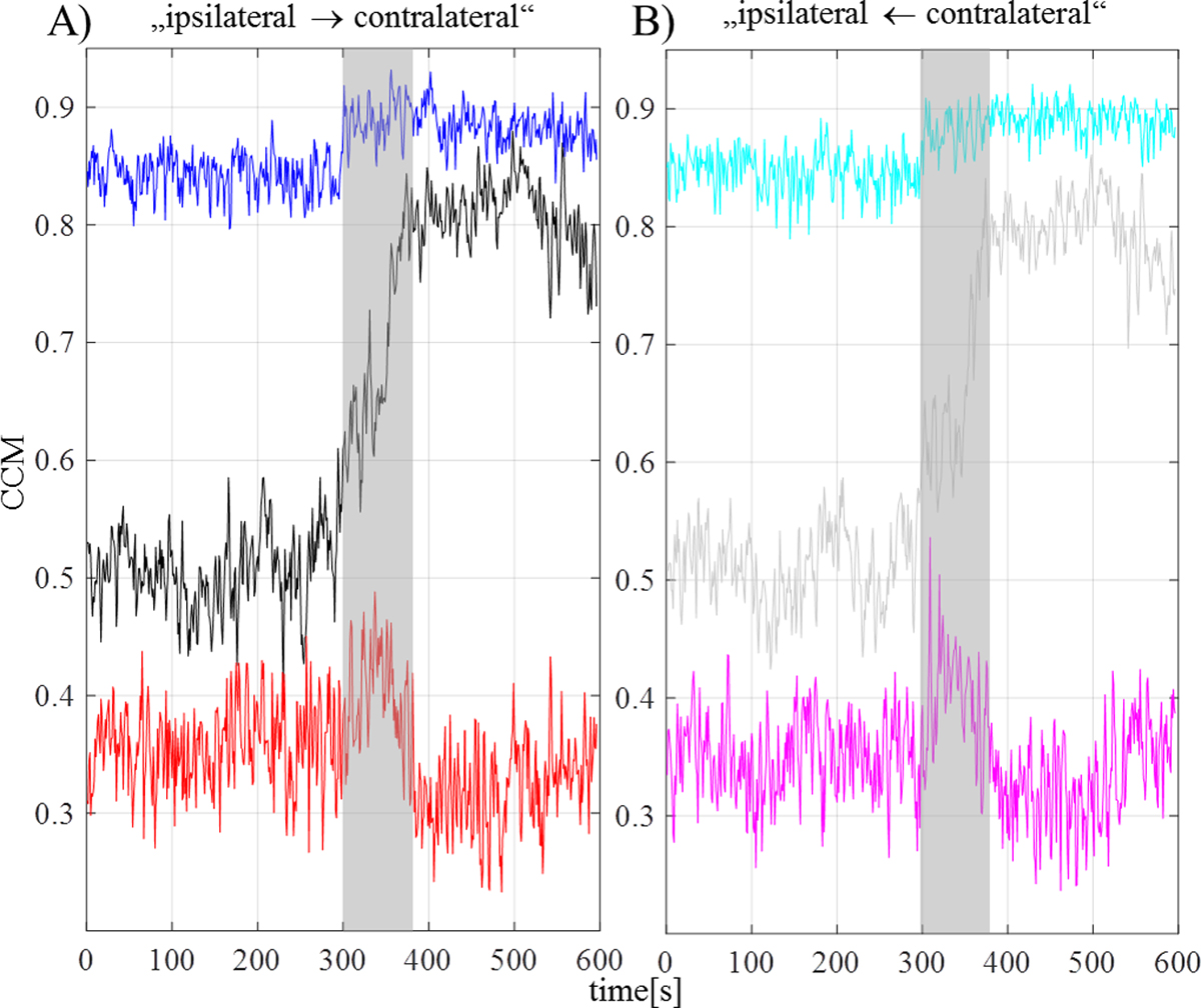
Download original image
Results of time-varying interval-based investigation of CCM of the ipsilateral vs. contralateral focus area. A sliding window of 4 s (=256 data points) is used. In (A) and (B), both directions of interval-based results of CCM of EEG activity (black and gray), δ-EEG activity (blue and cyan), as well as α-EEG activity (red and magenta) are shown (mean result of all children; color code of direction according to Fig. 3). Gray rectangle marks onset and median length of seizure.